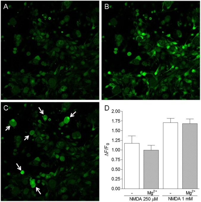
Fig. 4.
NMDA-induced calcium transients in satellite cells in the presence or absence of Mg2+. A–C show confocal images of Fluo-3 AM-loaded primary DRG cells cultured for 36 h, in which the satellite cells are observed to proliferate and migrate to the bottom of the dish. (A) Basal fluorescence of DRG cells. (B) Increase in fluorescence observed immediately after NMDA (250 μM) administration. (C) Increase in fluorescence observed in neurons after the administration of capsaicin (1 μM), used to verify the viability of nociceptive neurons. (D) Maximal fluorescence increase after NMDA (250 μM or 1 mM) administration in the presence or absence of Mg2+ (0.9 mM) in buffer. Results represent the means ± SEM of 17–22 cells in three different experiments. Statistical analysis, performed by using ANOVA followed by Bonferroni posttest, failed to detect significant differences between the groups.

