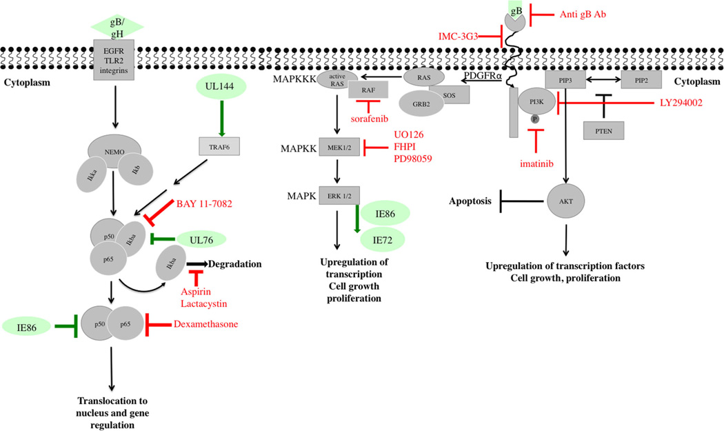
Figure 1. Modulation of PDGFR, MAPK and NF-κB pathways by CMV.
Depicted are schematics of PDGFR-α, MAPK and NF-κB signaling pathways as well as downstream effects and resulting cell activities. Viral proteins that modulate or interact with these pathways at various stages are shown in green. Compounds or antibodies that were reported to inhibit CMV replication by targeting specific steps in a given pathway are shown in red. Cellular proteins appear in gray. CMV, cytomegalovirus; MAPK, mitogen-activated protein kinase; NF-κB, nuclear factor kappa bet; PDGFR, platelet-derived growth factor receptor.

