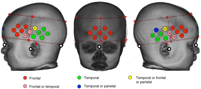
Fig. 4.
The fNIRS channels are projected onto a three-dimensional reconstruction of an infant. The red dashed lines and markers identify the position of the fNIRS headband on the infant head. For each fNIRS channel located within this headband, the identity of the underlying lobe (using the lobar atlas) is illustrated according to whether or not—when the channel was projected onto the cortical surface—over 75% of the group had a common region (frontal/temporal) or whether or not the identified region was split across the group with 30% to 60% in each of two to three lobes. The white markers indicate the position of the nasion, inion, and preauricular points on the infant head. Note that for the infants in the 4.5- and 6-month group all channels have the same majority identity accept for channel 5 and 25 (see Table 1).

