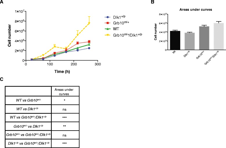
Figure 2.

Proliferation rates of E14.5 primary embryonic fibroblasts (PMEFs). A) Growth curves were plotted for E14.5 PMEFs seeded at equivalent densities and then cultured for 264 hours. Each data point represents the mean of three independent experiments (each comparing cells of all four genotypes), four replicates for each time point, counted twice. B) Areas under curves calculated using data from A. C) Statistical analysis of total areas under growth curves revealed that Grb10 m/+ and Grb10 m/+ /Dlk1 +/p PMEFs each proliferated significantly more than both wild type and Dlk1 +/p cells. All values represent means ± SEM, tested using one way ANOVA with Tukey’s post-hoc analysis; * P <0.05; ** P <0.01; *** P <0.001. For each genotype, n = 3. ANOVA, analysis of variance; E, embryonic day; SEM, standard error of the mean.
