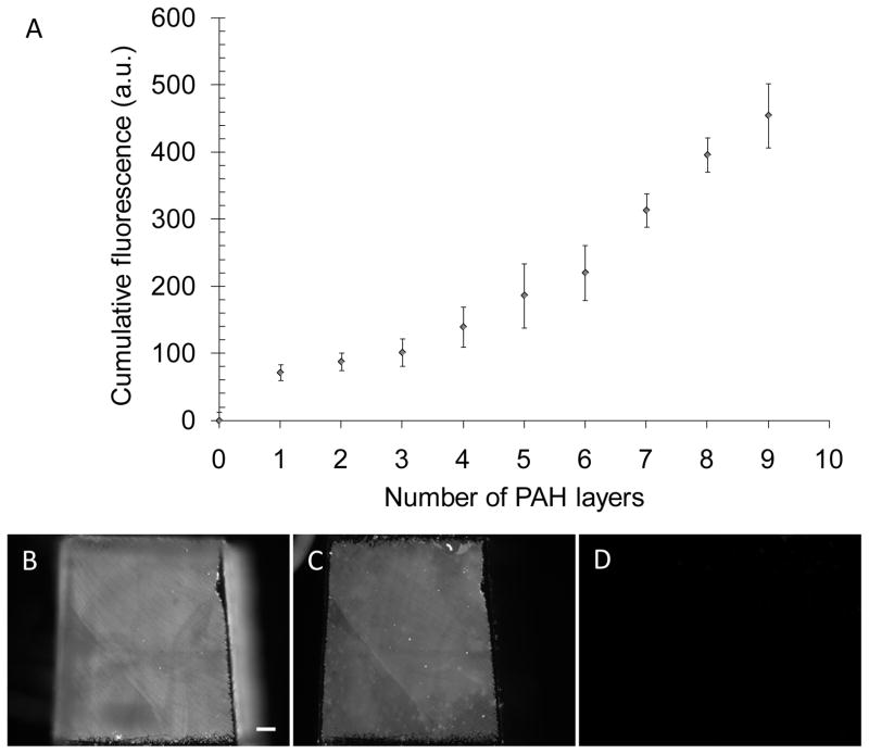
Figure 2.
Characterization of the fabrication of PEMs on PDMS stamps, and demonstration of the absence of contact transfer of the PEMs from the PDMS stamps onto skin-dermis. (A) Growth in the cumulative fluorescence of a (PAH/PAA)10 multilayers, containing FITC-labeled PAH, during fabrication on a PDMS substrate. (B, C, D) Micrographs of the fluorescent PEMs comprised of (PAH/PAA)10 on PDMS before the stamping, PDMS after stamping skin-dermis, and skin-dermis after the stamping, respectively. Scale bar=200 μm.

