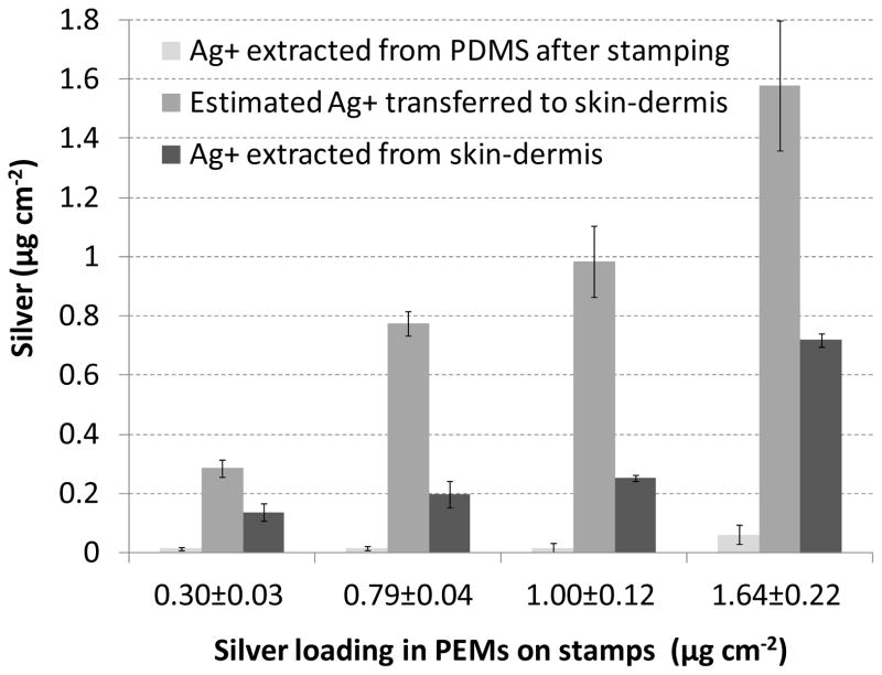
Figure 8.
Characterization of the transfer of silver onto skin-dermis by stamping with PEMs containing microspheres and a range of silver-loadings. The loading indicated under each set of bars is the silver in each PEM prior to stamping. The bars (left to right) show the amount of silver remaining on the PDMS after stamping skin-dermis; the amount of silver transferred onto skin-dermis (calculated as difference between the loading of silver extracted from PDMS before and after stamping); and the amount of silver extracted from stamped skin graft.

