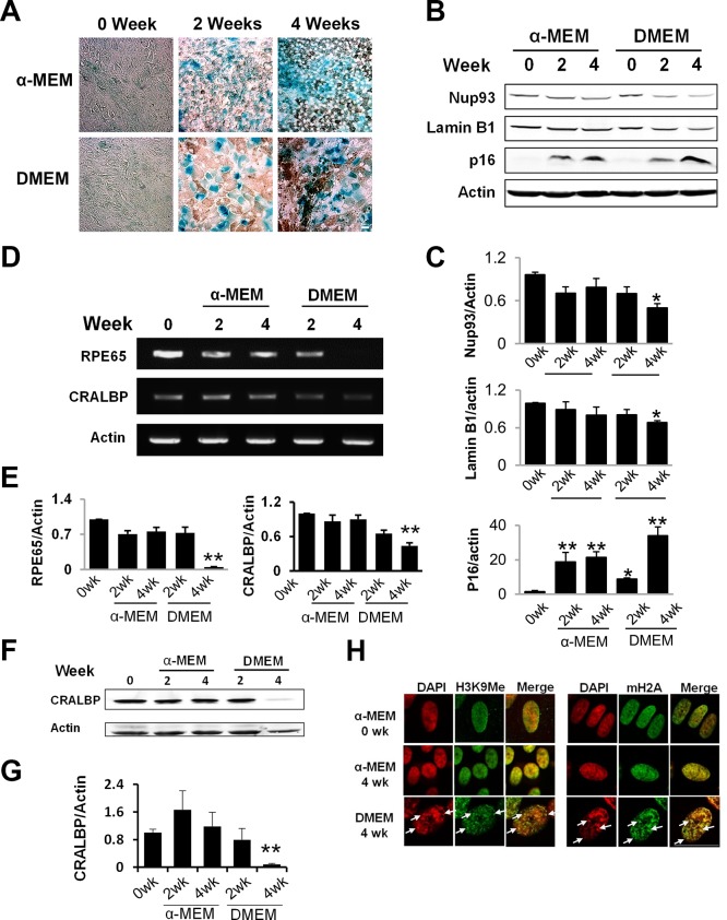
Figure 1.
Accelerated aging in postmitotic RPE cells cultured under suboptimal condition. (A) Confluent hfRPE cells were cultured in either α-MEM or DMEM culture medium for the indicated time. Activity of SA-β-gal was stained at pH 6.0. (B) Western blot analyses of nuclear envelope proteins. Data quantitation is presented in (C). (D) Reverse transcription–PCR analyses of RPE65 and CRALBP expression in cells at different stages of aging. Data quantitation is presented in (E). (F) Western blot analyses of CRALBP. (G) Quantitation of Western data. Data are the averages of three to five independent experiments (mean ± SEM; *P < 0.05; **P < 0.01, one-way ANOVA and Dunnett's post hoc test). (H) Immunofluorescent staining of heterochromatin foci (arrows), enriched with methylated histone H3 (H3K9Me) and MacroH2A.1. Scale bar: 20 μm.

