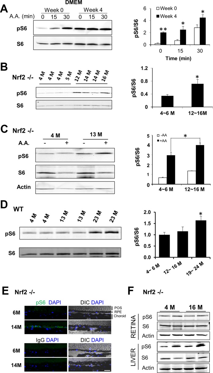
Figure 3.
Age-related changes of mTORC1 activity in the RPE, in vitro and in vivo. (A) Cultured RPE cells in DMEM medium for 4 weeks were stimulated with amino acid mixture for 15 or 30 minutes. The phosphorylation level of S6 was determined by Western blot analyses. Data presented are the averages of six independent experiments (mean ± SEM; *P < 0.05, **P < 0.01, Student's t-test). (B) Basal level of S6 phosphorylation in RPE/choroid tissues isolated from Nrf2-deficient mice at the indicated ages. (C) Ex vivo activation of mTORC1 in the RPE. Eyecups prepared from Nrf2-deficient mice at different ages were treated with amino acid mixture for 25 minutes. Activity of mTORC1 was assessed by the phosphorylation status of S6. Data presented are the averages of three separate experiments (*P < 0.05, Student's t-test). (D) Basal level of S6 phosphorylation in RPE/choroid tissues isolated from wild-type mice at the indicated ages. Data presented are the averages of four independent experiments (mean ± SEM) (*P < 0.05, one-way ANOVA, and Dunnett's post hoc test). (E) Immunostaining of phosphorylated S6 (green) on cryosections of posterior poles prepared from Nrf2-deficient mice at 6 and 14 months. Control IgG staining was presented in the lower panel. CH, choroid. Scale bar: 20 μm. (F) Activity of mTORC1 in liver or retina from young and old Nrf2-deficient mice.

