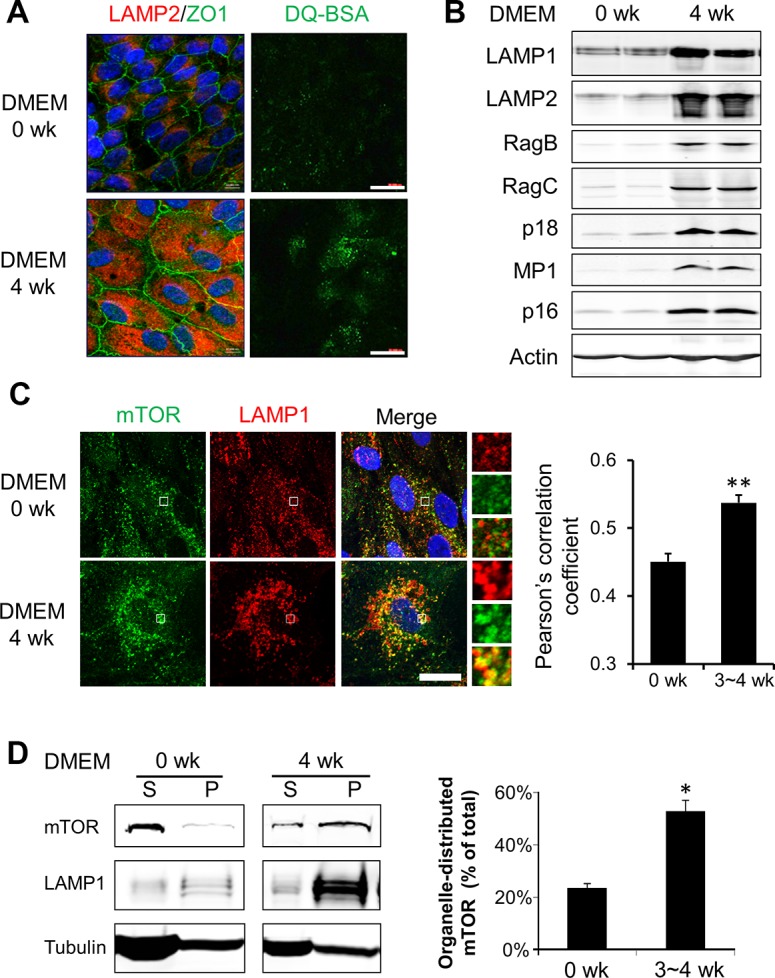
Figure 4.

Increased lysosome distribution of mTOR in aged RPE cells in vitro. (A) Left: Immunofluorescent staining of lysosome membrane protein LAMP2 and junction-associated protein ZO-1 in young and aged hfRPE cells. Right: live cell staining with lysosome probe DQ-BSA. Scale bar: 20 μm. (B) Western blot analyses of protein components of the Ragulator-Rag complex in cultured RPE cells. (C) Colocalization of mTOR (green) and LAMP1 (red) in young and aged RPE cells (0 and 3 ~ 4 weeks in DMEM culture medium, respectively). (Insets showed an enlarged field of interest). Pearson's correlation coefficient was calculated from images from >50 cells to quantify the colocalized signals. **P < 0.01, Student's t-test. (D) Western blot analyses of soluble (S) and organelle-associated (P) mTOR in young and aged RPE cells. Cells were permeabilized by saponin and fractionated into cytosolic (S) and organelle-enriched (P) fractions. Data presented are the averages of four separate experiments. *P < 0.05, Student's t-test.
