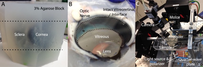
Figure 1.

Experimental approach. (A) Eyes are cleaned of orbital tissue and embedded in agarose. (B) Razorblades are used to section the block at the upper and lower margins of the cornea (dashed lines in [A]) providing an optically transparent view of the vitreous and an intact vitreoretinal interface (dashed region in [B]). (C) The sample is placed in a tissue bath for QPLI, as previously described.22 Further details describing this imaging technique are provided in the Methods section.
