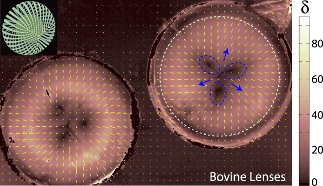
Figure 3.

Bovine lenses. Inset: Reference schematic for lens suture (yellow “Y”) and fiber cell organization (green) in the bovine lens (adapted from Kuszak JR, Zoltoski RK, Sivertson C. Fibre cell organization in crystalline lenses. Exp Eye Res. 2004;78(3):673–687. Copyright © 2003 Elsevier Science Ltd.). (Left, right) Two bovine lenses. The lens on the right is annotated. Fiber cells are oriented radially in all locations and retardation (δ) is substantially higher than vitreous samples. Y-shaped sutures are present (blue arrows) and regions of reduced retardation (dashed blue circles) are found between suture lines. Another zone of reduced retardation is located near the lens periphery (dashed white line). Both regions correspond to locations where fiber cells presumably curve out of the imaging plane.
