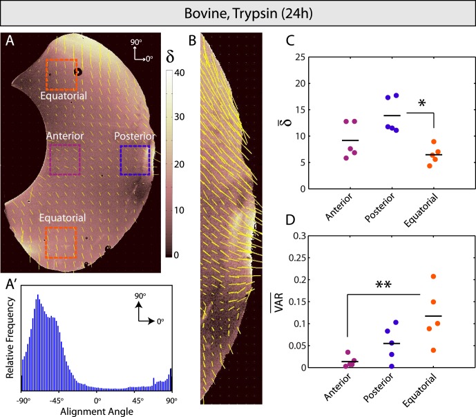
Figure 5.
Enzymatic vitreolysis with trypsin. (A, A′) Optical retardation (δ, indicating microscopic fiber alignment) is higher and the global orientation of remaining fibers is more homogeneous than bovine controls. (B) Fibers are aligned most strongly and in a transverse orientation to the vitreoretinal interface in the posterior vitreous. (C) Fibers are significantly more aligned in the posterior as compared to equatorial vitreous (P < 0.05). (D) Circular variance indicates significantly less anisotropy in the equatorial versus anterior vitreous (P < 0.01).

