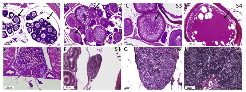
Fig. 6.
Ovarian and testicular stages in control stickleback fish. Panels A–D show ovarian follicle developmental stages S1–S4. Follicles representative of the stage are denoted with a black arrow. Panel E shows a representative undifferentiated gonad (U) in an adult (>300 dpf) male. Panels F–H show spermatogenic stage progression S1–S3. Staging schema condensed from Sokolowska and Kulczykowska (2006).

