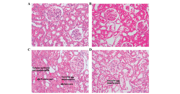
Figure 1.
Representative renal histomorphological changes in the (A) NN, normal diet group, (B) HN, high cholesterol diet group, (C) HL, high cholesterol plus low-osmolar contrast media iohexol group and (D) HLP, high cholesterol plus iohexol plus pentoxifylline group. (C) In iohexol-injected rats, tubular epithelial cell shedding and basement membrane nudity, vacuolar degeneration of tubular epithelial cells, protein cast, tubular dilation, loss of tubular brush border, and necrosis of partial tubular epithelial cells were observed. However, in (D) the rats treated with pentoxifylline and iohexol, only renal tubular epithelial cell degeneration was observed. Hematoxylin and eosin staining, original magnification, ×200.

