
Figure 5.
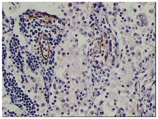
VEGFR-3 positive, small lymphatic vessels were observed in the peritumoral tissue with thin walls, discontinuous endothelial cells and interruptions of lymphocytes (magnification, ×100). A horseradish peroxidase-conjugated secondary antibody were used, and hematoxylin was used as a counterstain for the HRP-conjugated secondary antibody. VEGFR, vascular endothelial growth factor receptor.