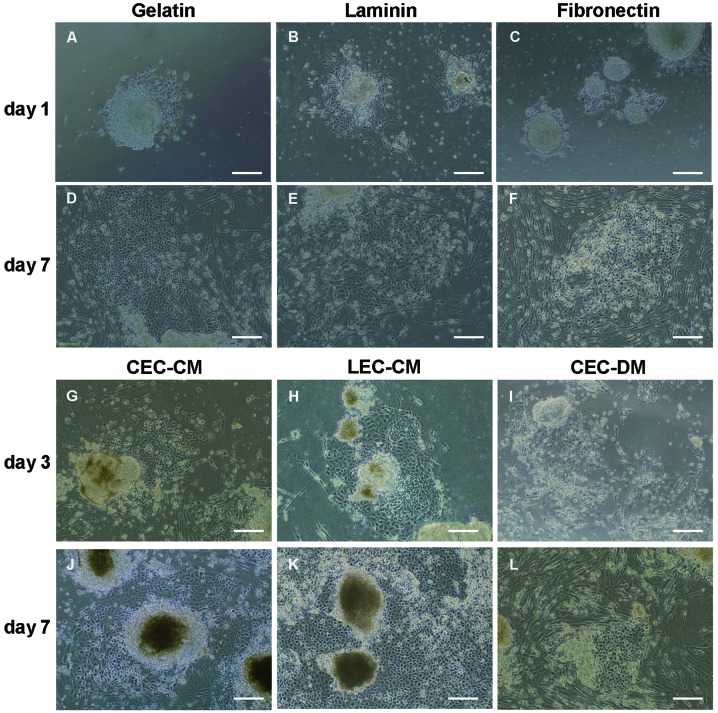
Figure 5.
Morphology of EB cell migration and differentiation in CM. Diverse effects of different coatings on EB cell migration and differentiation were found. During early migration on day 1 and late migration on day 7 after EB growth on various coatings: (A, D) gelatin (B, E) laminin, and (C, F) fibronectin. EBs continued to differentiate on gelatin-coated plates in CEC-CM and LEC-CM with CEC-DM as a control. Cells migrated out of the EBs and became endothelium-rich colonies. Induced pluripotent stem cells (G) in CEC-CM, (H) LEC-CM and (I) CEC-DM on day 3; (J) CEC-CM, (K) LEC-CM and (L) CEC-DM on day 7. Scale bars: A–L, 100 μm. EB, embryoid body; CM, conditioned medium; DM, differentiation medium; CEC, corneal endothelial cell; LEC, lens epithelial cell.

