
Figure 6.
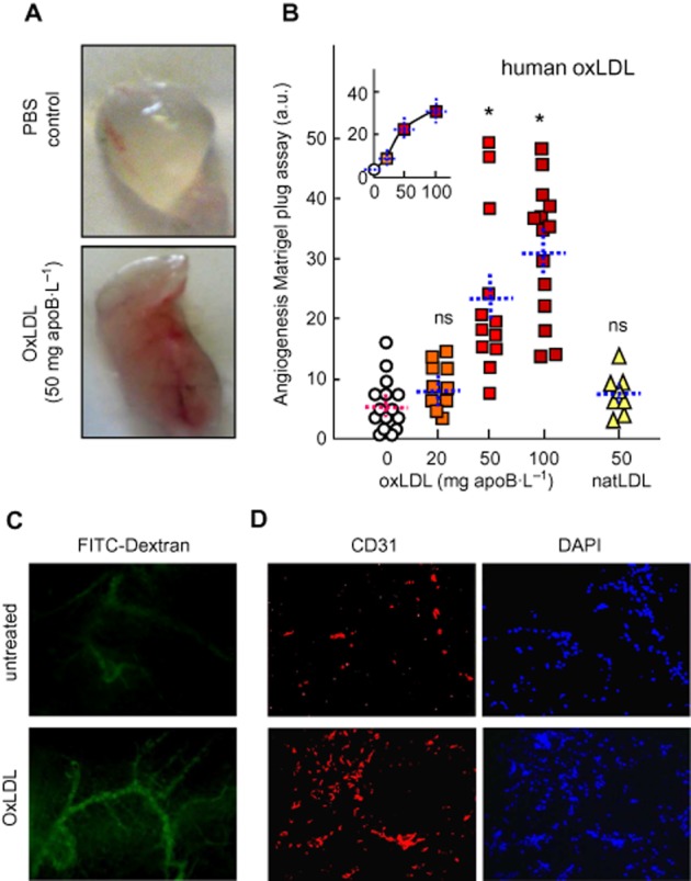
Human oxLDL stimulate angiogenesis in the murine Matrigel plug model. (A, B) C57BL/6 mice (8–14 per group), were injected s.c. with 0.4 mL Matrigel containing human native (natLDL) or oxLDL. Plugs were removed after 2 weeks and photographed. (A) Representative macrophotographs of plugs. Angiogenesis was evaluated by image analysis (Supporting information Figure S3 and S4). (B) Quantitative evaluation of angiogenesis in plugs containing PBS (circles), oxLDL (squares) or natLDL (triangles). Each point represents the value of angiogenesis in one plug. Means ± SEM are indicated by the dotted lines. Statistical analysis by one-way anova and Holm–Sidak (compared with PBS (0) group). *P < 0.05; ns, not significant. (C) Representative microphotographs of capillaries visualized by fluorescent FITC-dextran (mice injected i.v. with FITC-dextran 10 min before killing), and (D), plug sections labelled with anti-CD31 antibody and counterstained with DAPI.