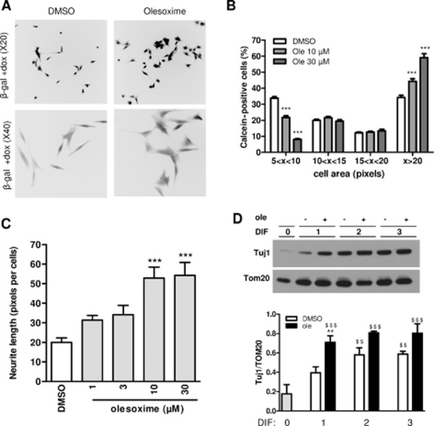
Figure 4.

Olesoxime accelerates β-gal SHSY-5Y differentiation and has a neurotrophic effect. β-gal-expressing SHSY-5Y cells seeded in 96-well plates at 3000 cells·cm−2 (low density) (A–C), or 30 000 cells·cm−2 (high density) (D) in the presence of doxycycline (dox), were differentiated with 10 μM RA 24 h after seeding and treated with olesoxime (ole) or DMSO at the same time. (A) Representative reverse black and white images of calcein-labelled neurons treated with DMSO or olesoxime (30 μM) were acquired using a fluorescence microscope (20× or 40× objective in the upper or lower figures respectively). (B) Calcein-labelled neurons in low-density cultures were individually counted with Trophos' Plate Runner HD after 5 days differentiation and cell distribution was classified according to cell body area. One representative experiment is shown. ***P < 0.001 compared with DMSO. (C) Neurite length per cell in olesoxime-treated cultures relative to DMSO controls was determined using images acquired using a fluorescence microscope (20x objective) and MetaMorph software. One representative experiment is shown. ***P < 0.001 compared with DMSO. (D) Expression of β-tubulin III using Tuj1 monoclonal antibody in differentiated SHSY-5Y cells treated with DMSO or olesoxime (30 μM) at different time points was monitored by immunoblotting. A representative experiment is shown and Tuj1-labelled protein levels were quantified using ImageJ and expressed relative to TOM20 protein levels. Mean ± SEM (n = 4). $$P < 0.01 $$$P < 0.001 compared with DIF0, **P < 0.05 compared with DMSO.
