Figure 1.
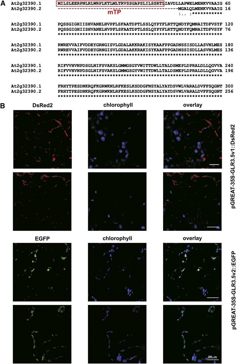
Splicing variants of AtGLR3.5. A, Alignment of N-terminal regions of the two AtGLR3.5 isoforms. Amino acid sequences are shown. The predicted mitochondrial targeting sequence (TargetP) is in the red box. According to ChloroP1.1, the chloroplast-targeting sequence is 82 amino acids long. B, Subcellular localization of the AtGLR3.5 isoforms with predicted mitochondrial (isoform 1) and chloroplast (isoform 2) targeting. Expression of pGREAT-2x35S-GLR3.5v1::DsRed2 (top) and pGREAT-2x35S-GLR3.5v2::EGFP (bottom) in 4-week-old Arabidopsis leaves is shown. The isoform1-DsRed2 fusion protein is located in highly motile structures resembling mitochondria (Supplemental Movies S1 and S2). Results shown are representative of three independent experiments giving the same results. Bars = 20 µm.

