Figure 4.
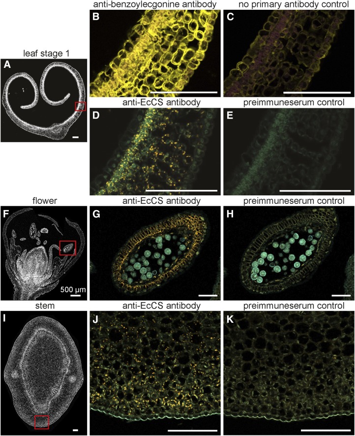
Immunolocalization of cocaine and cocaine synthase. Fluorescence micrographs of immunolabeled cross sections of different E. coca organs. A, Overview of L1 cross section with the region of interest marked by a red rectangle. B and C, L1 cross section immunolabeled with antibenzoylecgonine antibodies and no primary antibody, respectively. Fluorescence signal of secondary antibody shown in yellow. Background autofluorescence shown in purple. D and E, L1 cross section immunolabeled with polyclonal anticocaine synthase antibodies and preimmune serum, respectively. Fluorescence signal of secondary antibody shown in orange. Background autofluorescence shown in cyan. F, Overview of flower cross section with the region of interest marked by a red rectangle. G and H, Flower cross section immunolabeled with polyclonal anticocaine synthase antibodies and preimmune serum, respectively. Fluorescence signal of secondary antibody shown in orange. Background autofluorescence shown in cyan. I, Overview of stem cross section with the region of interest marked by a red rectangle. J and K, Stem cross section immunolabeled with polyclonal anticocaine synthase antibodies and preimmune serum, respectively. Fluorescence signal of secondary antibody shown in orange. Background autofluorescence shown in cyan. Single sections were probed with primary antibody (anticocaine, anticocaine synthase, or preimmune serum) and secondary antibody (anti-rabbit conjugated to HRP) and subsequently assayed with fluorescent tyramide substrate. Excitation of fluorophore for cocaine imaging was at 543 nm and detection using a 585- to 615-nm band-pass filter. Plant autofluorescence was excited at 488 nm and detected using a 505-nm low-pass filter. For imaging of cocaine synthase excitation of fluorophore was at 561 nm and detection using 585- to 614-nm channels. Plant autofluorescence was excited at 488 nm and detected using 495- to 534-nm channels. All pictures are overlays of fluorophore and autofluorescence channels. Bars = 100 µm (unless otherwise indicated).

