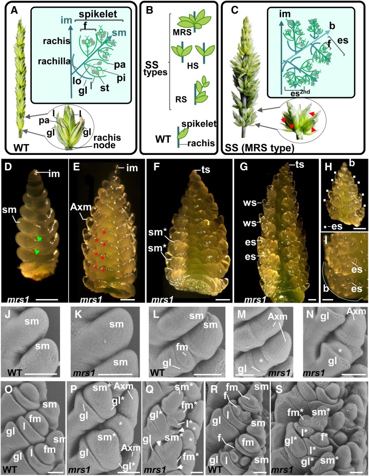
Figure 1.
SS phenotypes in bread wheat. A, Schematic representations of a spike (left) and a spikelet (right) from bread wheat N67 and of a theoretical wild-type (WT) spikelet (boxed). B, Schematic illustration of various SS structures: a cluster of spikelets at a rachis node referred to as a MRS, three spikelets (triple spikelet), and two spikelets in horizontal positions at a rachis node referred to as HSs; additional sessile spikelets with lateral branch bearing spikelets at a node referred to as a RS; and a single spikelet at a node for the wild-type spike. C, Illustration of the spike structure in the line NIL-mrs1 harboring numerous SSs (left), a rachis node with additional spikelets (right) where SSs are indicated with red arrows, and schematic representation of a branch-like structure (b) bearing ectopic spikelets at a rachis node in the lower part of the NIL-mrs1 spike (boxed). D to G, Light microscopy images of the NIL-mrs1 inflorescence at several developmental stages. D, Illustration of the spikelet differentiation stage; green arrows indicate glume primordia. E and F, Early floret differentiation stage. E, Secondary AxMs that later produce ectopic spikelets are indicated with asterisks. F, Spikelet meristems of ectopic spikelets form glume primordia. G, Late floret differentiation stage when all floral organs of wild-type spikelets (ws) are differentiated in the upper part of the inflorescence. H, A branch-like structure dissected from a rachis node of the NIL-mrs1 inflorescence. White squares indicate the ectopic spikelet (es). I, Location of ectopic spikelets at rachis nodes of the NIL-mrs1 inflorescence. Bars = 0.25 mm. J to S, Scanning electron microscopy images of N67 and NIL-mrs1 inflorescences at various developmental stages. J and K, Spikelet differentiation stage in the wild type (J) and mrs1 (K). L, Early floret differentiation stages when the spikelet meristem produces the FM in the wild type. M and N, Differentiation of secondary AxMs (indicated by asterisks) in the mrs1 mutant. O, Early floret differentiation stage showing lemmas. P and Q, The development of glumes (gl* and indicated by white arrows), lemma, and FMs by secondary AxMs (indicated by asterisks) in the mrs1 mutant. R, Floret differentiation stage showing differentiated floral organs in a basal floret of the wild type. S, The development of ectopic spikelets in mrs1. Bars =100 μm. es2nd, Ectopic spikelet of the second order, developing at the place of a floret of the first order ectopic spikelet; f , floret with floret organ primordia; f*, floret of an ectopic spikelet; Fm, floret meristem; Fm*, floret meristem of an ectopic spikelet; gl, glume; im, inflorescence meristem; l, lemma; l*, lemma of an ectopic spikelet; lo, lodicule; pa, palea; pi, pistil; ts, terminal spikelet; sm, spikelet meristem; sm*, spikelet meristem of an ectopic spikelet; st, stamen.

