
Figure 4. The procedural steps of in vivo experiment using VX2 rabbit liver tumor model.
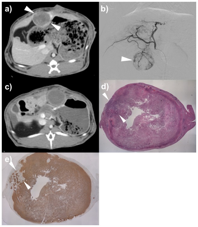
a) CT scan of portal venous phase showed a well-demarcated solitary tumor in the left hepatic lobe of the rabbit (arrowheads). b) Common hepatic angiography shows a hypervascular tumor staining (arrowhead). c) CT scan of portal venous phase 1 week after TACE using drug-eluting beads demonstrates no enhancement within the tumor (arrowheads). d) Photomicroscopic slide of the tumor specimen indicates focal viable tumor (arrowheads) (Hematoxylin-Eosin staining, X1). e) TUNEL staining indicates focal viable tumor (arrowheads).