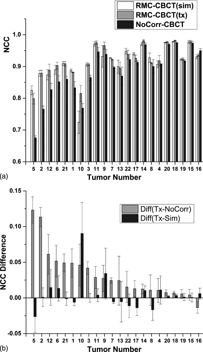
FIG. 7.
(a) NCC inside a VOI around the tumor in CBCT images before and after motion correction, following rigid registration with the gated CBCT. (b) Difference in NCC between RMC-CBCT(tx) and NoCorr CBCT, and between RMC-CBCT(tx) and RMC-CBCT(sim) CBCT. In both plots, tumor cases are sorted in order of decreasing difference in NCC (RMC-CBCT(tx) minus NoCorr). Column bars indicate mean NCC from five measurements; error bars indicate standard deviation.

