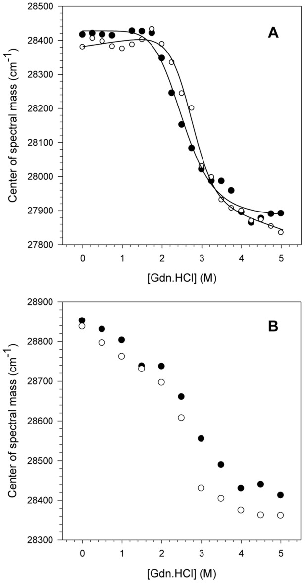
Figure 1. pH effects on the NS3 tertiary structure upon chemical denaturation.

The CM values obtained for NS3hel (A) and NS3FL (B) were calculated at pH 6.4 (closed circles) and 7.2 (open circles) upon increasing Gdn.HCl concentrations using Equation 1 (Material and Methods). The fluorescence spectra were obtained at 25°C and assay buffers were composed of 50 mM MOPS-NaOH (pH 6.4 or 7.2), 200 mM NaCl, 5 mM β-mercaptoethanol and 5% glycerol. The protein concentration was 1 µM.
