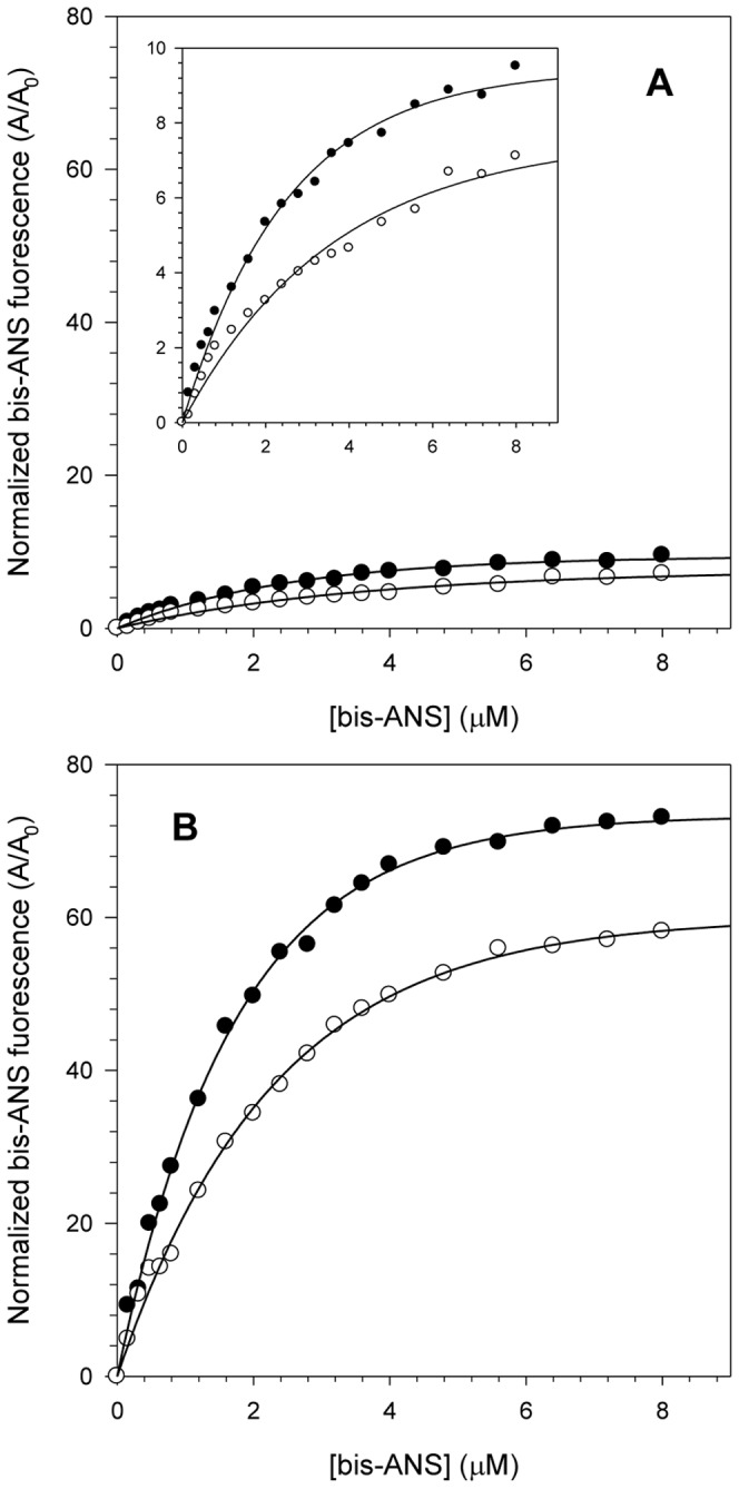
Figure 4. Interaction of the fluorescent extrinsic probe bis-ANS with NS3 at pH 6.4 and 7.2.

bis-ANS concentrations ranging from 0 to 8 µM were used to compare NS3hel (A) and NS3FL (B) hydrophobic clefts exposure at pH 6.4 (closed circles) and 7.2 (open circles). The inset in the graph A shows a reduction in the y-axis scale to demonstrate more clearly the effect of increasing bis-ANS fluorescence at both pH. Each point corresponds to the mean of the normalized bis-ANS fluorescence intensity obtained in three independent experiments. Spectra were acquired at 25°C in buffer solutions composed of 50 mM MOPS-NaOH (pH 6.4 or 7.2), 200 mM NaCl, 5 mM β-mercaptoethanol and 5% glycerol. The protein concentration was 1 µM.
