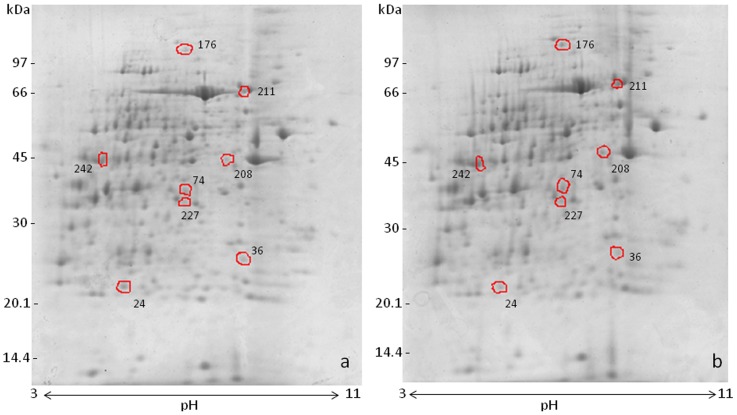
Figure 1. Two-dimensional gel electrophoresis of rat kidney lysate; Coomassie blue-stained gels from control (a) and MSG-treated rats (b).
Proteins were resolved on 7 cm pH 3–11 IEF strips (NL) followed by SDS-PAGE (12%). The differentially expressed spots detected by the image Master 2D Platinum 7.0 software are circled. The gels shown are representative of three independent experiments.

