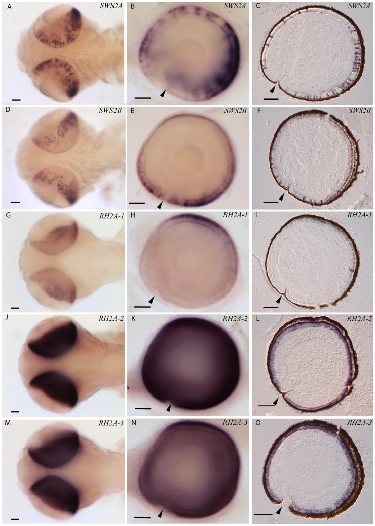
Figure 4. Topographic mRNA expression of visual opsins in cod larvae.
The retinal expression of different cone opsins were investigated by in situ hybridization on 18 days post fertilized cod larvae (2 days post hatching). The pictures in the left row (A, D, G, J, and M) show dorsal view of whole mount in situ on larvae, while the centre row of pictures (B, E, H, K and N) show whole mount of a right eye, lateral view; with larval anterior to the left. Sectional in situ is shown by sagittal sections in the left row of pictures (C, F, I, L and O). Expression of the various cone opsins was visualized by specific dig-labelled RNA probes: SWS2A (A, B and C), SWS2B (D, E and F), RH2A-1 (G, H and I), RH2A-2 (J, K and L) and RH2A-3 (M, N and O). Arrows indicate location of choroid fissure. Scale bars, 500 µm.

