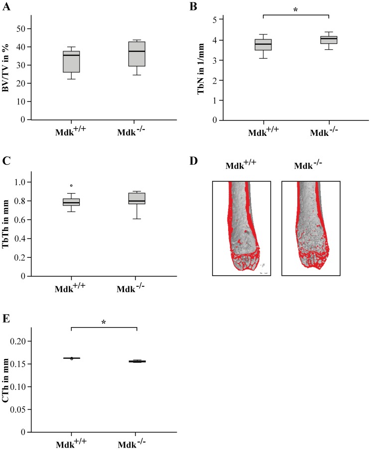
Figure 1. Mdk-deficient mice aged 9 months displayed increased trabecular number and decreased cortical thickness in the femur.
A) Trabecular bone volume to tissue volume ratio assessed by μCT analysis of volume of interest (VOI) 1 in the intact femur. B) Trabecular number assessed by μCT analysis of the VOI 1 in the intact femur. C) Trabecular thickness. D) Representative μCT images of the intact femurs. E) Cortical thickness assessed by μCT analysis of VOI 2 in the intact femur. *Significantly different from wildtype (p<0.05). (n = 6–7 per group).

