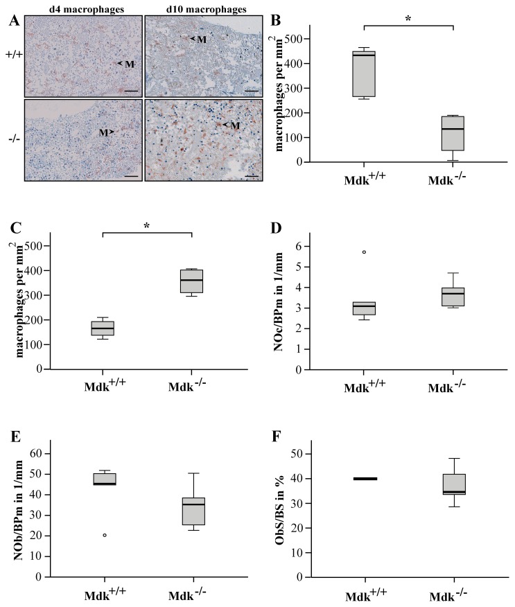
Figure 5. Presence of macrophages was delayed in Mdk-deficient mice.
A) Immunohistochemical staining for macrophages at days 4 and 10. Representative images showing recruited macrophages in the marrow cavities proximal to the osteotomy gap. M = macrophage; scale bar 50 µm; 200-fold magnification. The number of macrophages was counted in the marrow cavities close to the osteotomy gap B) at day 4 and C) at day 10. D) TRAP-stained sections from fractured femurs were analyzed for the number of osteoclasts per bone perimeter. E) Toluidine-blue-stained sections were analyzed for the number of osteoblasts per bone perimeter and F) Osteoblast surface per bone surface. (n = 5–6 per group).

