Fig. 1.
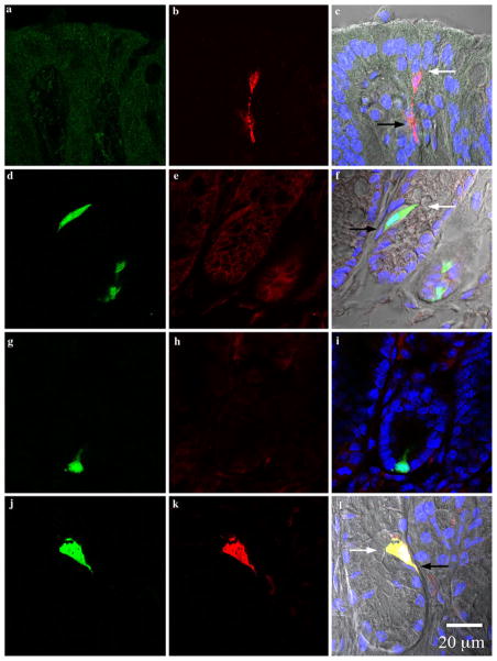
Photomicrographs from colonic crypts. All sections are aligned vertically with the lumen on top. White and black arrows point to the lumen and lamina propria, respectively. Colors represent the following: green green fluorescent protein (GFP), red PYY, blue DAPI, and gray differential interference contrast (DIC) and are merged from the left and middle columns. a–c Sections from wild type Swiss Webster mice stained for GFP (a) and PYY (b). GFP is absent and PYY cells appeared to have a spindle-like shape in the colon of wild type mice. d–f Sections from PYY-GFP mice incubated with secondary antibodies alone (DyLight488-conjugated donkey anti-chicken and Cy3-conjugated donkey anti-rabbit). Colonic cells from transgenic PYY-GFP mice expressed endogenous GFP and non-specific binding of secondary antibodies was absent. g–i Sections from a PYY-GFP mouse incubated in the presence of chicken anti-GFP and rabbit anti-PYY pre-adsorbed in excess (10 μM) of PYY peptide. The specificity of the antibody to PYY is shown by the absence of immunoreactivy after pre-absorption with synthetic PYY. j–l PYY-GFP sections incubated in the presence of chicken anti-GFP and rabbit anti-PYY antibodies. PYY immunoreactivity with the antibody exclusively co-localized with GFP positive cells (l, yellow). Please refer to online version of the article for colored figures

