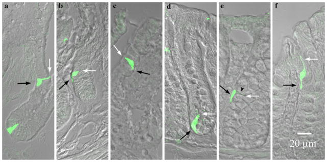
Fig. 4.

PYY-GFP cell morphology in the ileum versus colon. White and black arrows indicate the lumen and lamina propria respectively. a–c In the ileum a cells were columnar or flask-shaped at the bottom of the crypts and pseudopod-like basal processes were absent. Further up the crypt (b) towards the villus tip (c) basal processes became evident. d–f In the colon, flask-shaped cells were evident at the bottom of the crypts (d). A neck-like projection (black arrow head) and a basal process were evident in the cell in the middle of the crypt (e). Towards the apex of the crypt, this neck-like projection extended to maintain contact with the lumen, while the basal process remained in contact with the lamina propria (f). Note the sigmoidal appearance of the cell to maintain contact with the lumen as well as the lamina propria
