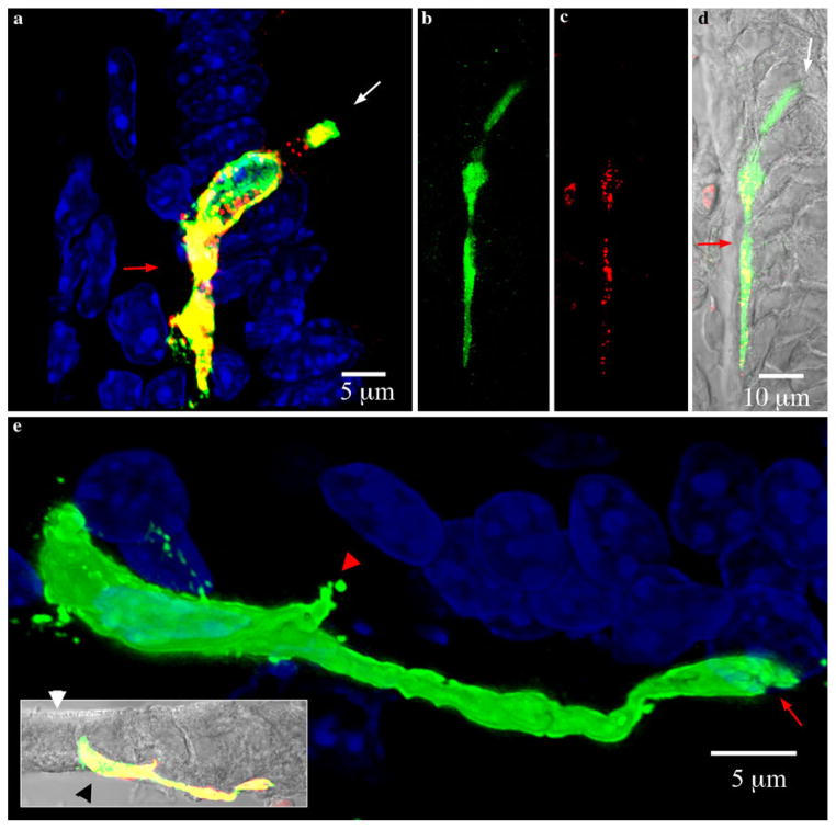
Fig. 5.
Pseudopod-like basal processes in PYY-GFP cells of the ileum. White and red arrows indicate the lumen and lamina propria respectively. Sections imaged were 15 μm thick. a Maximal projection view of typical PYY-GFP cell in a villus of the ileum. The pseudopod-like process runs on the basal side, in between the lamina propria and the base of other epithelial cells. The basal process typically extends downward towards the bottom of the crypt. b–d PYY-GFP cell with a long basal process extending along the base of adjacent epithelial cells. The total length of the cell is greater than 70 μm and the basal process is approximately 50 μm long. b Green GFP, c red PYY, d composite with DIC (gray). e Maximal projection view of PYY-GFP cell in a villus from the ileum. Inset shows the cell position with respect to the brush border (white arrow head) and lamina propria (black arrow head) (green GFP, red PYY staining, yellow merged images, gray DIC). The pseudopod-like basal process often ended in a bifurcation (red arrow). There are some smaller projections (red arrow head) that extended from the body of the cell

