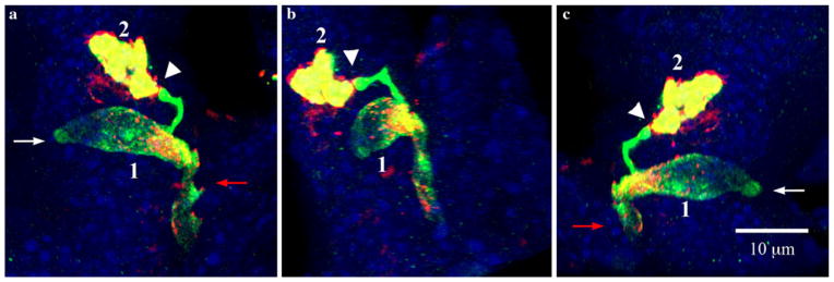
Fig. 6.

Relationship of a PYY-GFP cell with another PYY-GFP cell through synapse-like structures at the end of pseudopod-like processes. White and red arrows indicate the lumen and lamina propria respectively. Section imaged was 15 μm thick. a–c Views of three-dimensionally reconstructed PYY-GFP cells in a villus of the ileum from different angles. The synapse-like projection at the end of pseudopod-like process from cell 1 that makes contact with cell 2 is shown by the white arrowhead. Green GFP, red PYY staining, yellow merged images, blue DAPI
