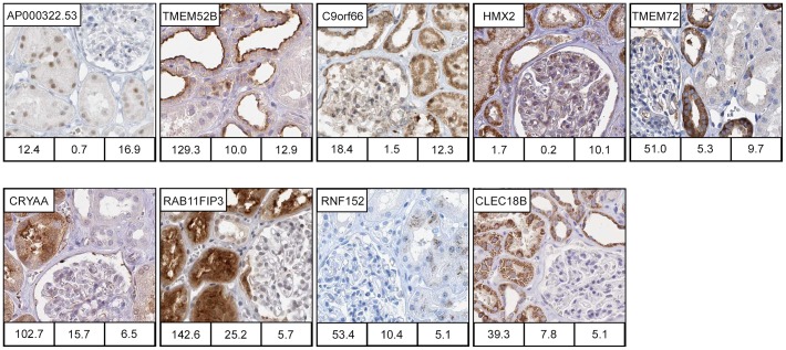
Figure 5. Kidney enriched proteins previously not described in kidney.
Immunohisto-chemically stained images of kidney enriched proteins (TS>5 FPKM) for which we found no previous description expression in kidney based searches in literatures or online databases (GeneCards, WikiGene, BioGPS). Of the 16 kidney enriched genes not described previously, IHC images confirming kidney specific localisation were available for 10 genes. In proximal tubule cells, AP000322.53 localise to the nucleus while TMEM52B, RAB11FIP3 and CRYAA show a general cytoplasmic staining, and RNF152 shows a granular cytoplasmic staining. In distal tubule, TMEM72 localises to the basolateral membrane. C9orf66 is found in all nephron segments but with different localisations. In glomeruli, it localise to the nucleus, in proximal tubular cells to nucleus and cytoplasm, while in cells in distal tubule and collecting duct only in cytoplasm. Both HMX2 and CLEC18B localises to the cytoplasm of cells in proximal and distal tubule, and also in the collecting duct, however for CLEC18B only in the intercalated cells. Numbers under each IHC image correspond to Mean FPKM in kidney (left bottom), Max FPKM in the tissue with 2nd most highly expressed level (middle bottom), and Tissue specificity score in kidney (right bottom).

