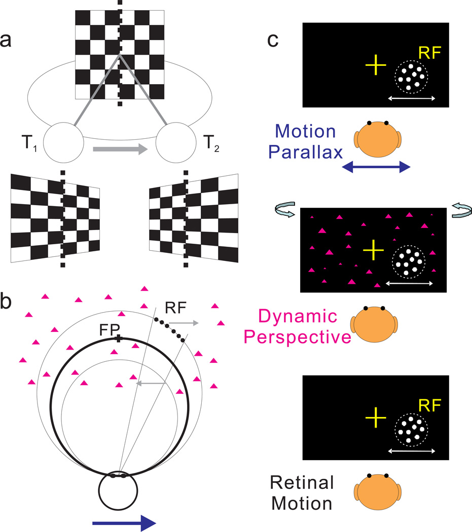
Figure 1. Schematic illustration of dynamic perspective cues and stimuli for measuring depth tuning from motion parallax.
(a) An observer translates from left (at time T1) to right (at time T2) while the observer’s eye (circle) rotates to maintain fixation at the center of a world-fixed checkerboard. As the observer translates, the perspective distortion changes dynamically and is manifest as a rotation of the image (in stimulus coordinates) about a vertical axis through the fixation target (dotted line). Supplementary Movie 1 shows an image sequence resulting from this viewing geometry. (b) Top view illustrating the stimulus geometry. The thick circle represents the locus of points in space for which motion simulates a depth equivalent to zero binocular disparity. The other two circles represent sets of points that have image motion consistent with particular near or far depths (equivalent to −1° and +1° of binocular disparity). Dots within the receptive field (which have no size cues to depth) are shown as filled black circles; background dots (having size cues) are shown as magenta triangles. When the observer moves rightward (blue arrow), near and far dots move in opposite directions (gray arrows). (c) Frontal views for each experimental condition. In the Motion Parallax condition, animals experience full-body translation and make counteractive eye movements to maintain fixation on a world-fixed target (yellow cross). In the Retinal Motion condition, the animal’s head and eyes are stationary, but visual stimuli replicate the image motion experienced in the Motion Parallax condition. The Dynamic Perspective condition is the same as the Retinal Motion condition except that a 3D cloud of background dots was added to the display. Background dots near the receptive field were masked.

