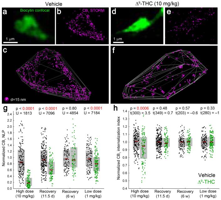
Figure 6. Chronic THC treatment induces long-lasting loss of CB1 from the plasma membrane of GABAergic axon terminals in a dose-dependent manner.
(a,d) Deconvolved confocal images of axon terminals of perisomatic interneurons, recorded in slices prepared from mice treated with vehicle or THC (i.p. injections twice a day for 6.5 days). (b,e) STORM imaging reveals a dramatic THC-evoked reduction of CB1 localization points. A CB1 antibody with improved sensitivity derived from transgenic rabbits (Online Methods) was used to enable reliable visualization of CB1 receptors at low and high levels alike. (c,f) Visualization of CB1 localization points in 3D demonstrates an increased intraterminal accumulation of CB1 in THC-treated animals. (g) Upon chronic administration of 10 mg/kg THC, CB1 NLP are reduced by 74% (n = 185 and 117 axon terminals from 3 vehicle- and 2 THC-treated animals, Mann-Whitney U test). After a drug-free period of 11.5 days, 53% of CB1 receptors recovered, but the reduction in NLP (35%) was still significant (n = 283 and 113 boutons from 3 and 3 animals). A longer recovery period of six weeks, however, allowed complete rescue (n = 113 and 92 boutons from 2 and 3 animals). Chronic treatment with a lower (1 mg/kg) dose resulted in a slight (16%) decrease in NLP (n = 129 and 153 boutons from 2 and 3 animals). (h) The remaining CB1 localization points are shifted towards the center of the axon terminal after high dose of THC (unpaired two-sided t-test). However, internalization was not detectable after recovery for 11.5 days or 6 weeks, or after low dose of THC. Graphs show raw data and median±IQR.

