
Figure 3.
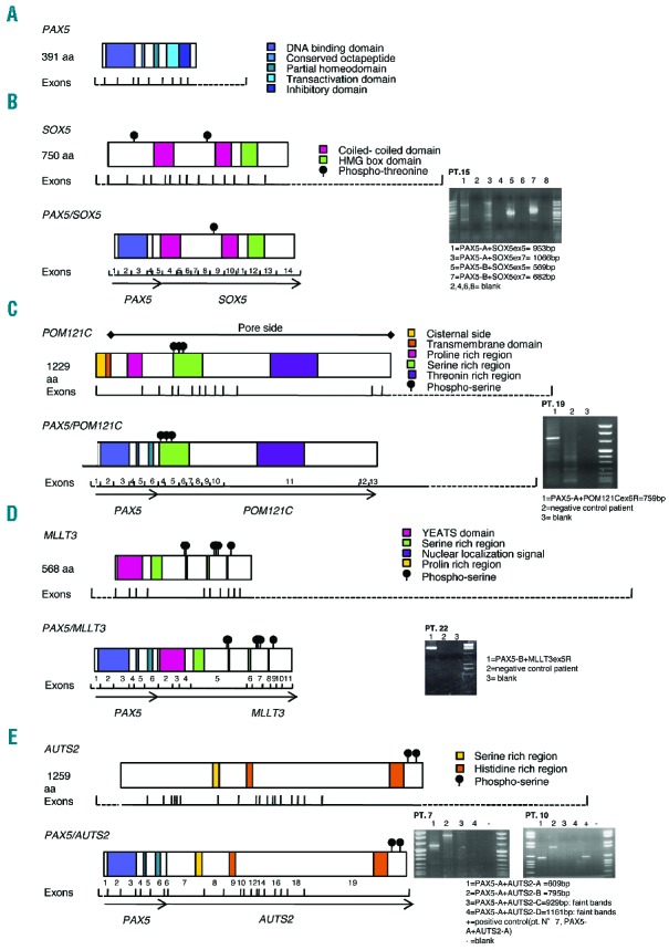
Schematic representation of novel PAX5 fusion proteins and mRNA expression levels. For each novel fusion gene (B–E), the RT-PCR gel electrophoresis (the specific primers reported in the Online Supplementary Table S4 are indicated) and the scheme of the predicted fusion protein are represented on the right and on the left, respectively. The conserved functional domains of the wild-type protein counterparts and exon numbers of fused transcripts are also indicated. (A) Normal PAX5. (B) PAX5/SOX5 in Patient 15. (C) PAX5/POM121C in Patient 19. (D) PAX5/MLLT3 in Patient 20. (E) PAX5/AUTS2 in Patients 7 and 10.