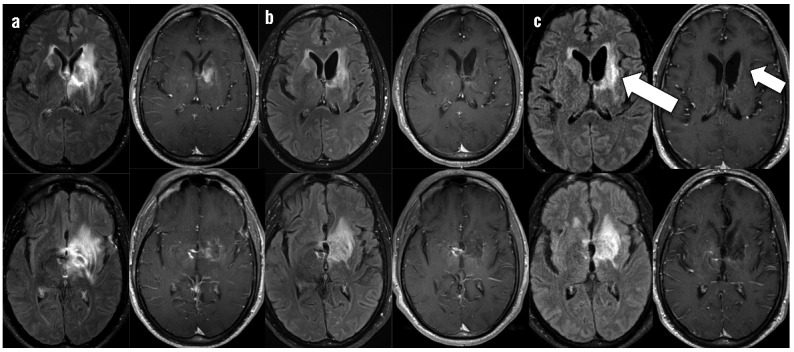
Figure 1.

Brain MRI post-contrast fluid attenuated inversion recovery (FLAIR) and post-contrast T1 sequences throughout clinical course. For time points a–c, left column shows FLAIR post-gadolinium axial images and right column shows T1 post-gadolinium axial images at the same level. a) Base-line contrast ring-enhancing mass in the left basal ganglia with surrounding edema prior to initiation of immunochemotherapy, b) Improvement in edema and enhancement prior to initiating the 3rd cycle of immunochemotherapy, T1 post-contrast images shows no residual ring enhancement. c) New progression of enhancement (left arrow) seen at cycle 3 day 24 when the patient became obtunded, T1 post-contrast image shows no recurrence of primary tumor despite new enhancement (right arrow).
