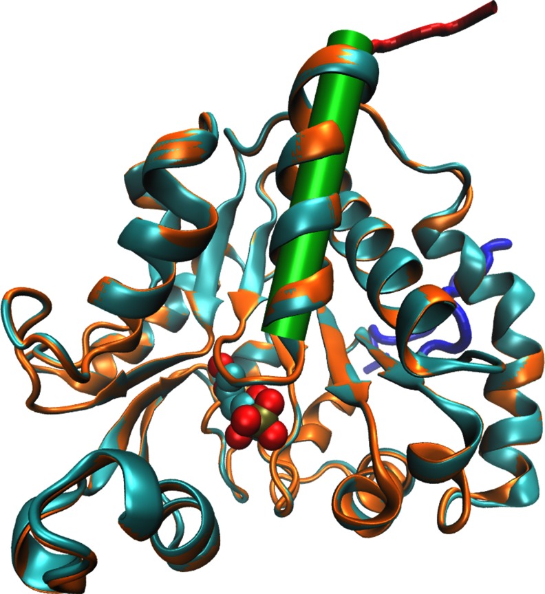
Fig. 2.
Superimposed model structures of GlmNagB-HisC (orange) and GlmNagB-HisN (cyan) with their additional oligo-His fragments, colored red and blue, respectively. The substrate F6P (transferred from the template structure 2bkx) is shown as a spacefill model bound in the active site. Location of the α1 helix is indicated by a green cylinder

