Figure 1.

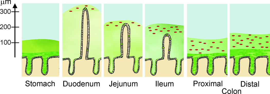
Schematic representation of the gastrointestinal mucus system. The mucus is depicted in green and the bacteria represented as red dots. The axis to the left shows the thickness of the mucus as measured in mice (8).

Schematic representation of the gastrointestinal mucus system. The mucus is depicted in green and the bacteria represented as red dots. The axis to the left shows the thickness of the mucus as measured in mice (8).