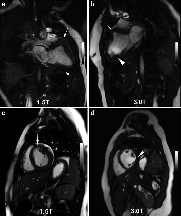
Fig. 1.
SSFP cine frames at 1.5 T (a, c) and 3.0 T (b, d) in the same boy with a low-grade, isointense intramyocardial tumor (white arrows), manifest as an aneurysm of the basal anteroseptal wall. Images were obtained three years apart at ages 1.5 years (6.8 kg, 1.5 T) and 4.9 years (16.3 kg, 3.0 T). Image quality was scored as fair at 1.5 T with mild off-resonance banding artifact (white arrowhead, a). At 3.0 T, the off-resonance artifact is more severe (white arrowhead, b, d) but did not render the images nondiagnostic

