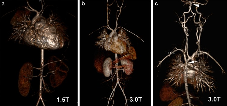
Fig. 5.
HR-CEMRA with volume-rendered reconstruction at 1.5 T (a) and 3.0 T (b) in the same 18-month-old (6.8 kg) boy with intramyocardial tumor as shown in Fig. 1. HR-CEMRA with volume-rendered reconstruction (c) at 3.0 T in the same 5-year-old boy (23.5 kg) whose SSFP cine is shown in Fig. 2. Note the basilar tip cerebral aneurysm (white arrowhead) and aortic coarctation (white arrow) distal to the left subclavian artery (LSA)

