
Figure 1.
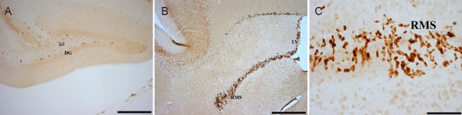
Representative photomicrograph of Ki-67 immunohistochemical staining in the various brain regions of four-striped mice.
(A) DG; (B) subventricular zone and RMS. (C) A magnified image of the RMS showing the darkly stained neurons. Scale bars: 20 μm in A, 10 μm in B and 1 μm in C. DG: Dentate gyrus; RMS: rostral migratory stream; hil: hilum; LV: lateral ventricle.