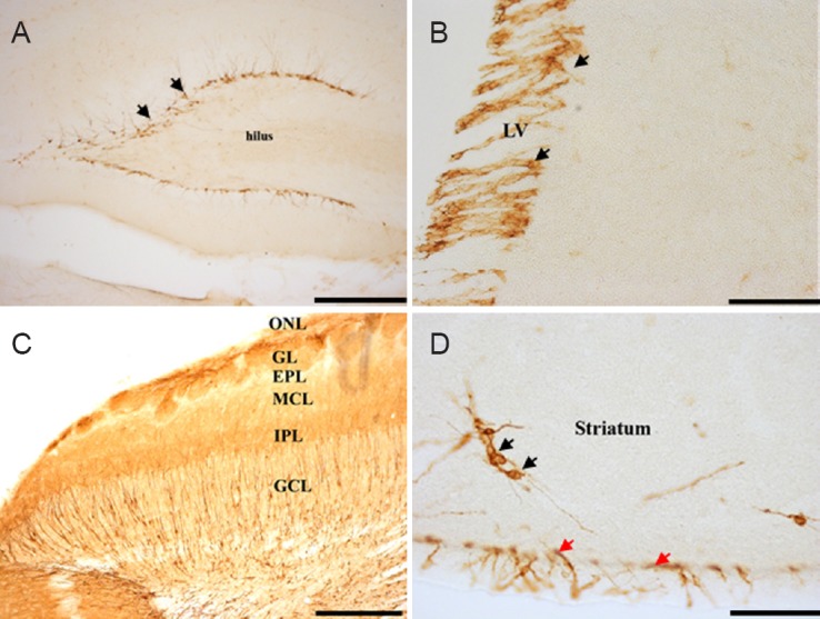
Figure 2.

Representative photomicrograph of doublecortin (DCX) immunohistochemical staining in the brain of four-striped mice.
(A) Dentate gyrus with the immunoreactive cells lining the whole length. (B) DCX-immunoreactive cells on the subventricular zone of the lateral ventricle. (C) Six different layers in the olfactory bulb region with the immature neurons. (D) The striatum and isolated neurons (black arrows) and the subventricular zone (red arrows). Scale bars: A, C, 20 μm; B, D, 2.5 μm. LV: Lateral ventricle; ONL: olfactory nerve layer; GL: glomerular layer; EPL: external plexiform layer; MCL: mitral cell layer; IPL: internal plexiform layer; GCL: granule cell layer.
