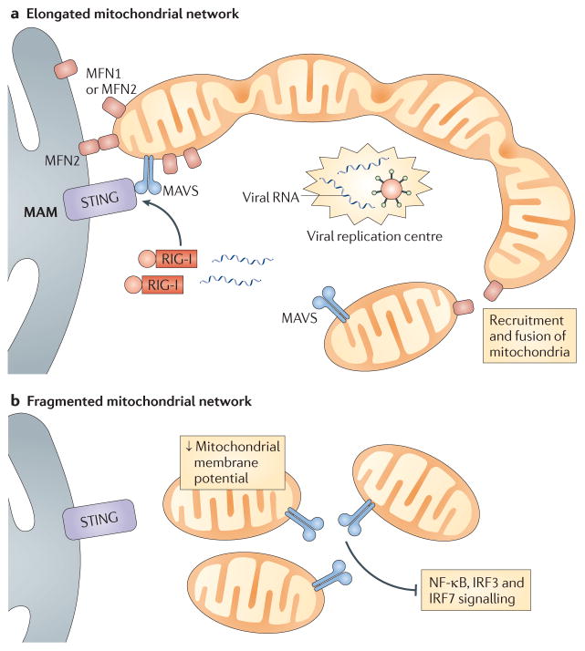
Figure 2. Mitochondrial dynamics regulate MAVS signalling.
a | During infection, retinoic acid-inducible gene I (RIG-I) and mitochondrial antiviral signalling protein (MAVS)-enriched mitochondria are recruited around centres of viral replication to promote MAVS signalling. This occurs as mitofusin 1 (MFN1) and MFN2 induce fusion of the mitochondrial network, which also serves to increase MAVS interactions with downstream signalling molecules. Mitochondrial MFN1 and MFN2 also interact with endoplasmic reticulum-localized MFN2, which promotes interactions between MAVS and stimulator of interferon genes (STING) at mitochondria-associated membranes (MAMs). b | Fragmentation of the mitochondrial network — which is induced by viral infection, mitofusin deficiency or overexpression of fission-promoting molecules — results in decreased mitochondrial membrane potential and blocks interactions between MAVS and signalling molecules such as STING. This leads to reduced signalling by nuclear factor-κB (NF-κB), interferon regulatory factor 3 (IRF3) and IRF7.

