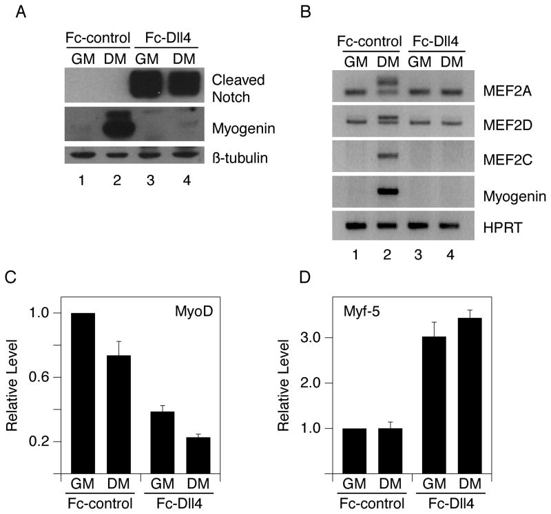
Figure 1.
Ligand-induced Notch signaling blocks myogenesis. 6-well plates were coated with 4.5 ml of ligand-containing supernatant per well. C2C12 cells were grown on Fc-Dll4-coated or Fc-control-coated plates and switched from growth medium (GM) to differentiation medium (DM) as indicated. Cells were analyzed after 24 hours for A) cleaved Notch1 and Myogenin proteins (Western); B) MEF2A, MEF2D, MEF2C and Myogenin RNAs (RT-PCR); C) MyoD and D) Myf-5 RNAs (quantitative RT-PCR). MyoD and Myf-5 levels shown in (C–D) are normalized to the Fc-control-GM condition (defined as 1) and plotted as the average +/− standard deviation of two replicate samples. The upper bands of the MEF2A and MEF2D RNA doublets are the differentiation-induced splice variants. β-tubulin protein and HPRT or 18S RNA were used as loading controls.

