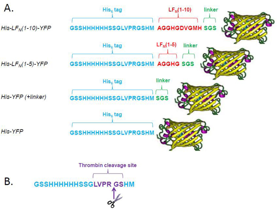
Figure 2.
A. Schematic of selected truncated His-LFN-YFP constructs. The N-terminal 20-residue His6-tag is shown in blue, LFN residues are shown in red, linker residues are shown in green, and the C-terminal YFP is shown as a yellow β-barrel. B. 20-residue His6-tag. Note that the N-terminal methionine residue encoded by pET-15b is not present, as it is removed during protein expression25. The site of thrombin cleavage is shown in purple, with the cleavage taking place between the arginine and glycine residues. Thrombin treatment removes the first 16 residues of the tag, leaving behind four residues (Gly-Ser-His-Met).

