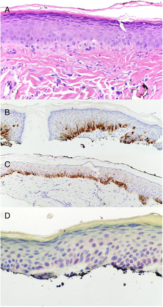
Fig 2.
R21 has a benign staining pattern in melanocytic lesions overlying a cicatrix. A) Hematoxylin and eosin staining of fibrous papule revealing an increased density of enlarged basal layer melanocytes (40×). B) Melan-A, brown stain, reveals increased density of melanocytes approaching confluence (20×). C) HMB45, brown stain, reveals increased density of melanocytes with rare cells above the basal layer in an area of irritation (20×). D) R21 (sAC), red stain, reveals perinuclear dot (benign) staining in melanocytes (40×).

