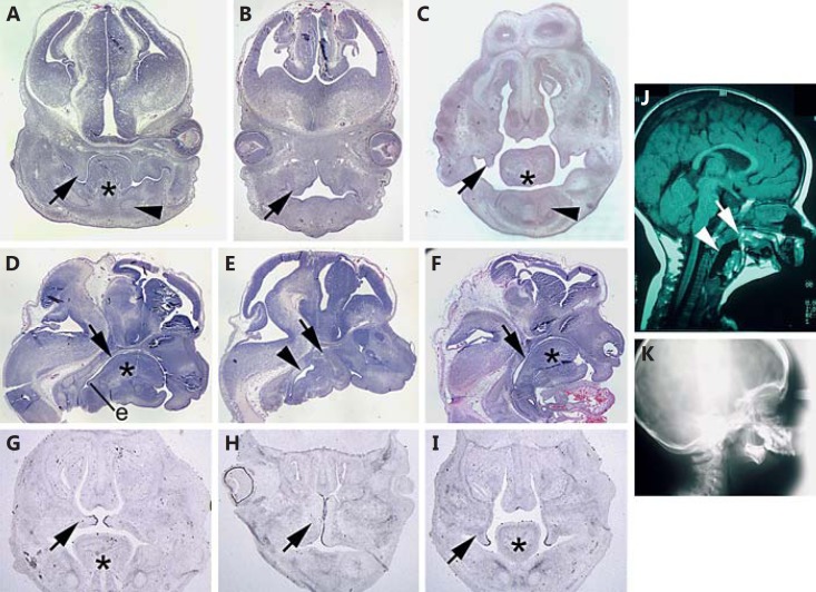
Fig. 3.
Oropharyngeal malformation in E13.5 snoopy embryos. In control E13.5 embryos (A), the vertical palatal shelves (arrow) are closely associated with the tongue, and Meckel's cartilage can be seen in cross section (arrowhead). In contrast, E13.5 snoopy mutants (B) fail to form a tongue, and the palatal shelves (arrow) are distended and abnormal in morphology. C In a Crklsno/sno embryo in which the tongue has formed, the palatal shelves take on a more typical morphology but are delayed in their elevation (arrow), and Meckel's cartilage is apparent (arrowhead). A sagittal section through the head of a control embryo (D) reveals the relationship of the tongue to the oral cavity. The oral cavity (arrow) is patent down to the epiglottis (e) and beyond into the larynx and opening of the oesophagus. A similar section in a snoopy embryo (E) demonstrates that there is no clear oral cavity and that the mandibular rudiment is continuous with the skull base (arrow). The pharynx distal to the occluded oral cavity (arrowhead) is typically distended relative to controls. A sagittal section through a mildly affected Crklsno/sno embryo (F) reveals mandibular hypoplasia and microglossia (asterisk) as well as a patent oral cavity (arrow). In situ hybridisation for Tgfb3 at E14.5 highlights the medial edge epithelium in the palatal shelves (arrow) of a wild-type embryo (G), in the dysmorphic palatal shelves of a severely affected Crklsno/sno embryo (H) or in the relatively normal but delayed palatal shelves of a mildly affected Crklsno/sno embryo (I). J Mid-sagittal computed tomography image of an individual with ACS illustrating a similar phenotype to snoopy including fusion of the mandible to the upper oropharynx (arrow) and a distended pharynx distal to the occlusion (arrowhead). K Lateral X-ray illustrating a diminutive mandible analogous to that observed in snoopy embryos. Asterisks indicate the tongue.

