Fig. 3.

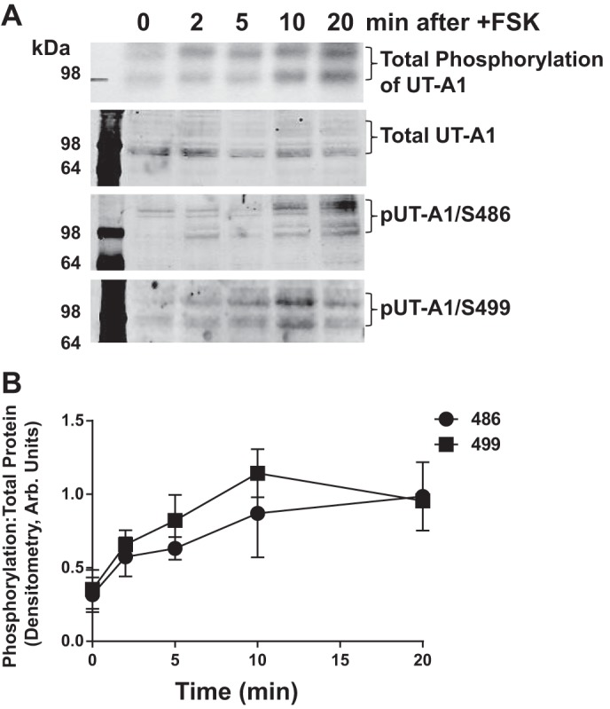
Phosphorylation of UT-A1 at S486 and S499 occur on the same time scale as total phosphorylation. Inner medullas were dissected from five Sprague Dawley rats, minced, and pooled together. Tissue was incubated in phosphate-free media containing [32P]orthophosphate (0.1 mCi/ml) for 2 h at 37°C. After tissues were loaded with phosphate, FSK (10 μM) was added to the pooled tissue, and equal volumes of tissue were collected and lysed at the indicated time points. Next, UT-A1 was immunoprecipitated. Autoradiography was used to determine total phosphorylation of UT-A1 (top). Western blot analysis was performed on the remaining samples, and immunoblot analysis was performed for total UT-A1, phosphorylation of UT-A1 at S486 (pUT-A1/S486), and phosphorylation of UT-A1 at S499 (pUT-A1/S499). A: representative autoradiographic images and Western blots, where brackets indicate the multiple glycosylation forms of UT-A1 observed in the tissue. B: time course of phosphorylation. Values are means ± SE; n = 5.
